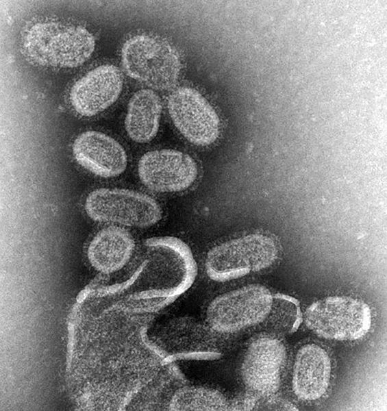
檔案:EM of influenza virus.jpg

Yi-lám thai séu: 565 × 600 chhiong-su. Khì-thâ kié-sak-thu: 226 × 240 chhiong-su | 452 × 480 chhiong-su | 700 × 743 chhiong-su.
Ngièn-pún tóng-on (700 × 743 chhiong-su, vùn-khien thai-séu: 82 KB, MIME lui-hîn: image/jpeg)
Vùn-khien li̍t-sṳ́
Tiám-khim ngit-khì / sṳ̀-kiên lòi chhà-khon tông-sṳ̀ chhut-hien-ko ke vùn-khien.
| Ngit khì / Sṳ̀-kiên | Suk-lio̍k-thù | Vì-thu | Yung-fu | Yi-kien | |
|---|---|---|---|---|---|
| tông-chhièn | 2007年8月10日 (Ńg) 13:41 |  | 700 × 743(82 KB) | ToNToNi | {{Information |Description=CDC, CDC Public Health Image Library (PHIL), http://phil.cdc.gov/Phil/details.asp |Source=Originally from [http://en.wikipedia.org en.wikipedia]; description page is/was [http://en.wikipedia.org/w/index.php?title=Image%3AEM_of_i |
Vùn-khien yung-chhú
Hâ poi ke 1-chak ya̍p-mien lièn-chiap to pún vùn-khien:
Chhiòn-vet tóng-on sṳ́-yung chhong-khóng
Hâ-lie̍t khì-thâ Wiki chûng sṳ́-yung liá-chak tóng on:
- af.wikipedia.org ke sṳ́-yung chhong-khóng
- an.wikipedia.org ke sṳ́-yung chhong-khóng
- ar.wikipedia.org ke sṳ́-yung chhong-khóng
- as.wikipedia.org ke sṳ́-yung chhong-khóng
- awa.wikipedia.org ke sṳ́-yung chhong-khóng
- azb.wikipedia.org ke sṳ́-yung chhong-khóng
- az.wikipedia.org ke sṳ́-yung chhong-khóng
- bat-smg.wikipedia.org ke sṳ́-yung chhong-khóng
- ba.wikipedia.org ke sṳ́-yung chhong-khóng
- be-tarask.wikipedia.org ke sṳ́-yung chhong-khóng
- be.wikipedia.org ke sṳ́-yung chhong-khóng
- bg.wikipedia.org ke sṳ́-yung chhong-khóng
- bn.wikipedia.org ke sṳ́-yung chhong-khóng
- bo.wikipedia.org ke sṳ́-yung chhong-khóng
- br.wikipedia.org ke sṳ́-yung chhong-khóng
- bs.wikipedia.org ke sṳ́-yung chhong-khóng
- bxr.wikipedia.org ke sṳ́-yung chhong-khóng
- ca.wikipedia.org ke sṳ́-yung chhong-khóng
- cdo.wikipedia.org ke sṳ́-yung chhong-khóng
- ckb.wikipedia.org ke sṳ́-yung chhong-khóng
- csb.wikipedia.org ke sṳ́-yung chhong-khóng
- cs.wikipedia.org ke sṳ́-yung chhong-khóng
- da.wikipedia.org ke sṳ́-yung chhong-khóng
- de.wikipedia.org ke sṳ́-yung chhong-khóng
- en.wikipedia.org ke sṳ́-yung chhong-khóng
- Influenza A virus
- Emergent virus
- Portal:Medicine/Selected Article Archive
- Wikipedia:Today's featured article/January 2007
- Wikipedia:Today's featured article/January 1, 2007
- Portal:Medicine/Selected article/8, 2008
- Portal:Medicine/Selected Article
- Portal:Medicine/Selected Article/10
- Influenza
- Wikipedia:VideoWiki/Influenza
- User:JenOttawa/Notes/practice
- User:Mr. Ibrahem/Influenza
- en.wikibooks.org ke sṳ́-yung chhong-khóng
- en.wikinews.org ke sṳ́-yung chhong-khóng
- et.wikipedia.org ke sṳ́-yung chhong-khóng
- eu.wikipedia.org ke sṳ́-yung chhong-khóng
- fa.wikipedia.org ke sṳ́-yung chhong-khóng
Kiám-sṳ liá vùn-khien ke kiên-tô chhiòn-vet sṳ́-yung chhong-khóng.

